Scanning Electron Microscopy
Search from Scanning Electron Microscopy stock photos, pictures and royalty-free images from iStock. Find high-quality stock photos that you won't find anywhere else.
Variable Pressure Scanning Electron Microscope Center for Biotechnology University of
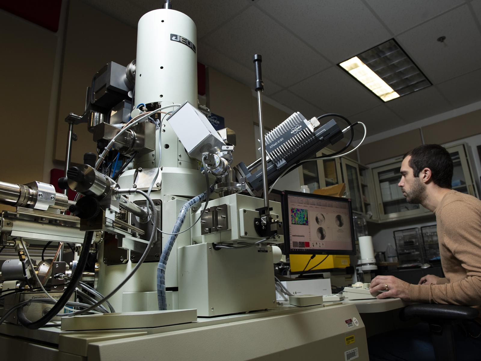
Browse 1,889 scanning_electron_microscope photos and images available, or start a new search to explore more photos and images. female scientist working with scanning electron microscope - scanning_electron_microscope stock pictures, royalty-free photos & images. salt crystal - scanning_electron_microscope stock pictures, royalty-free photos.
Scanning Electron Microscopy Gallery Center for Microscopy and Imaging
Scanning electron microscope photo- micrograph of an epon cast of a zoiJ of SiHiiliii'ont. Photographed X -00.. Please note that these images are extracted from scanned page images that may have been digitally enhanced for readability - coloration and appearance of these illustrations may not perfectly resemble the original work.

Scanning electron microscope (SEM) Definition, Images, Uses, Advantages, & Facts Britannica
1) Improving the quality of secondary electron images 2) Obtaining infromation different form that obtained when the specimen is not tilted, that is, observing topographic features and observing specimen sides. 3) Obtaining stereo micrographs. a) Dependence of image quality on tilt angle Fig. 13 shows a photo taken at a tilt angle of 0° (a.
Scanning Electron Microscopes (SEM) Adelaide Microscopy University of Adelaide
History. An account of the early history of scanning electron microscopy has been presented by McMullan. Although Max Knoll produced a photo with a 50 mm object-field-width showing channeling contrast by the use of an electron beam scanner, it was Manfred von Ardenne who in 1937 invented a microscope with high resolution by scanning a very small raster with a demagnified and finely focused.
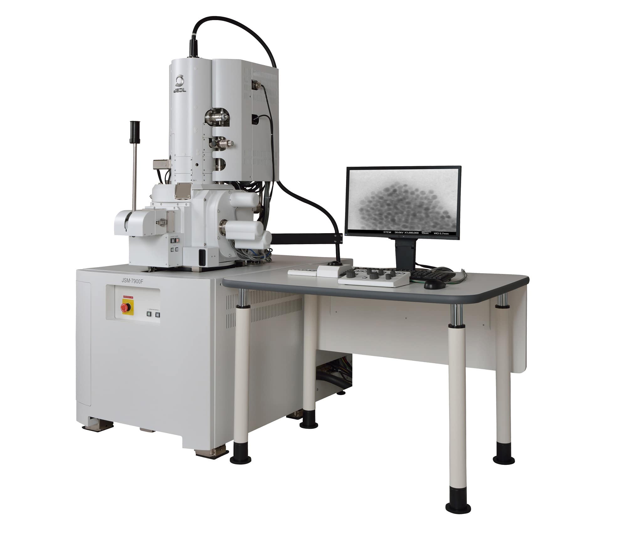
JEOL JSM 7001F/TTLS LV Scanning Electron Microscope PNNL
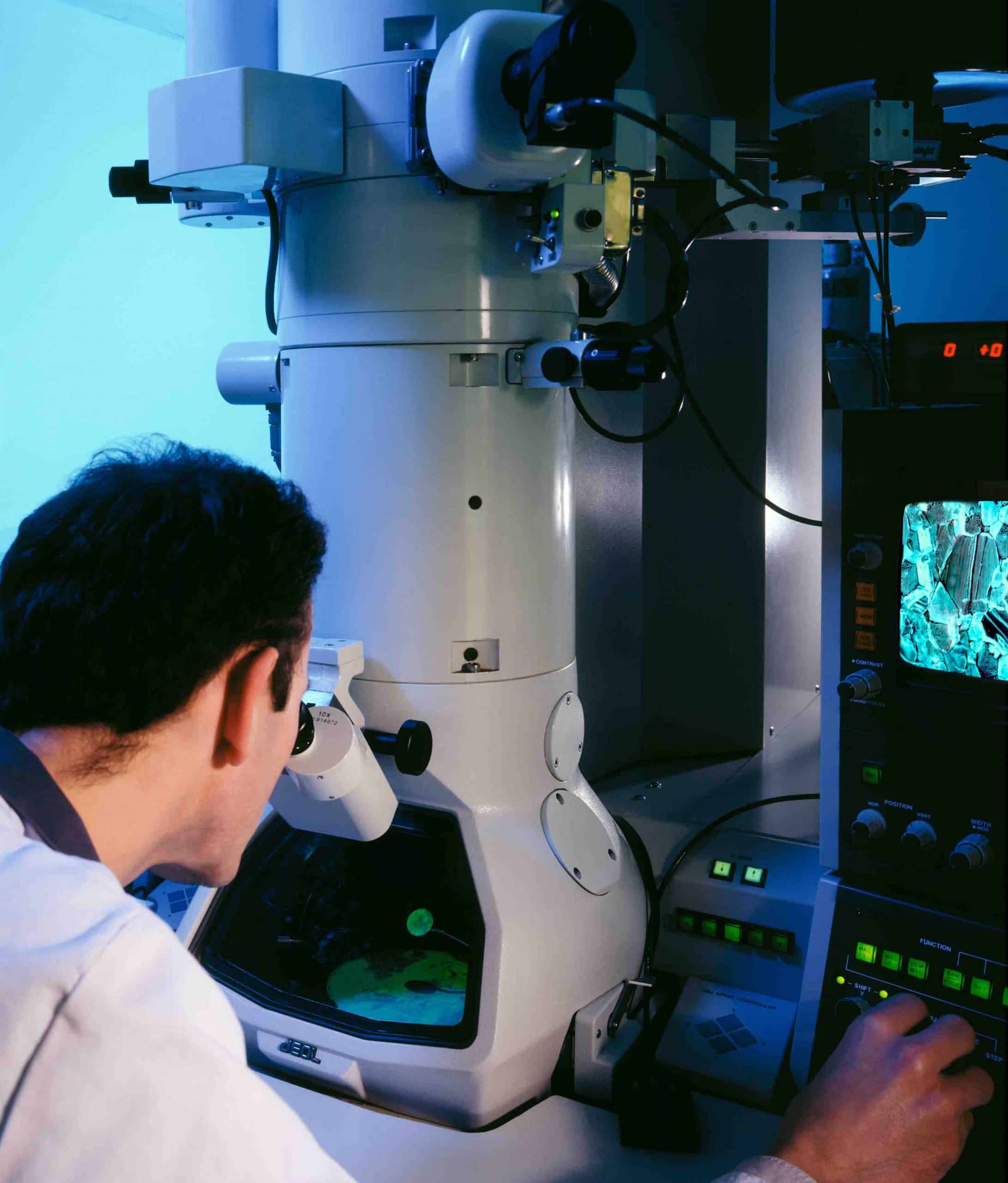
2,058 Scanning Electron Microscope Stock Photos and High-res Pictures. scanning electron microscope stock photos, high-res images, and pictures, or explore additional sem biology stock images to find the right photo at the right size and resolution for your project. field emission electron microscope in laboratory - scanning electron microscope.
Scanning Electron Microscope Pomona College in Claremont, California Pomona College
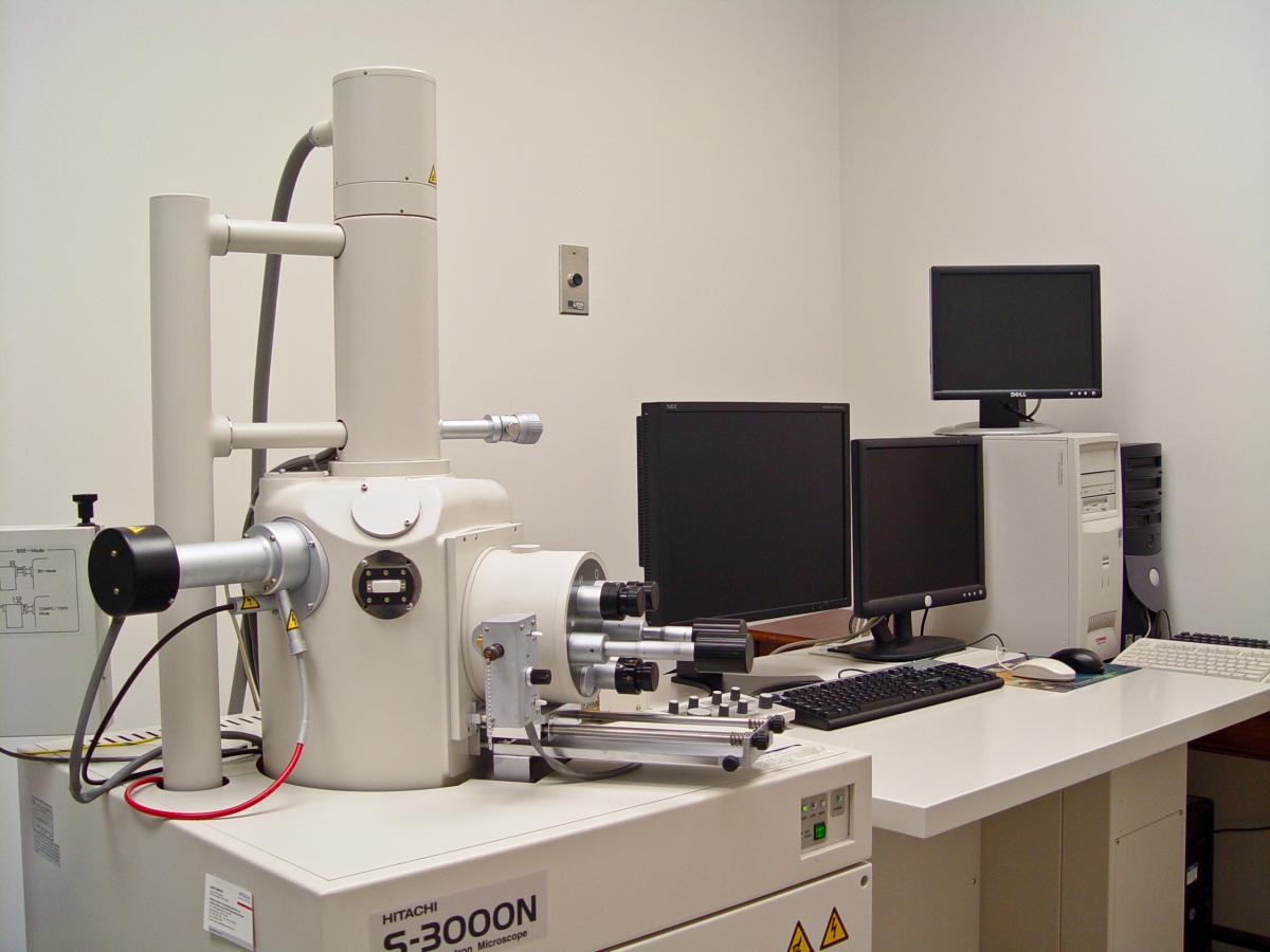
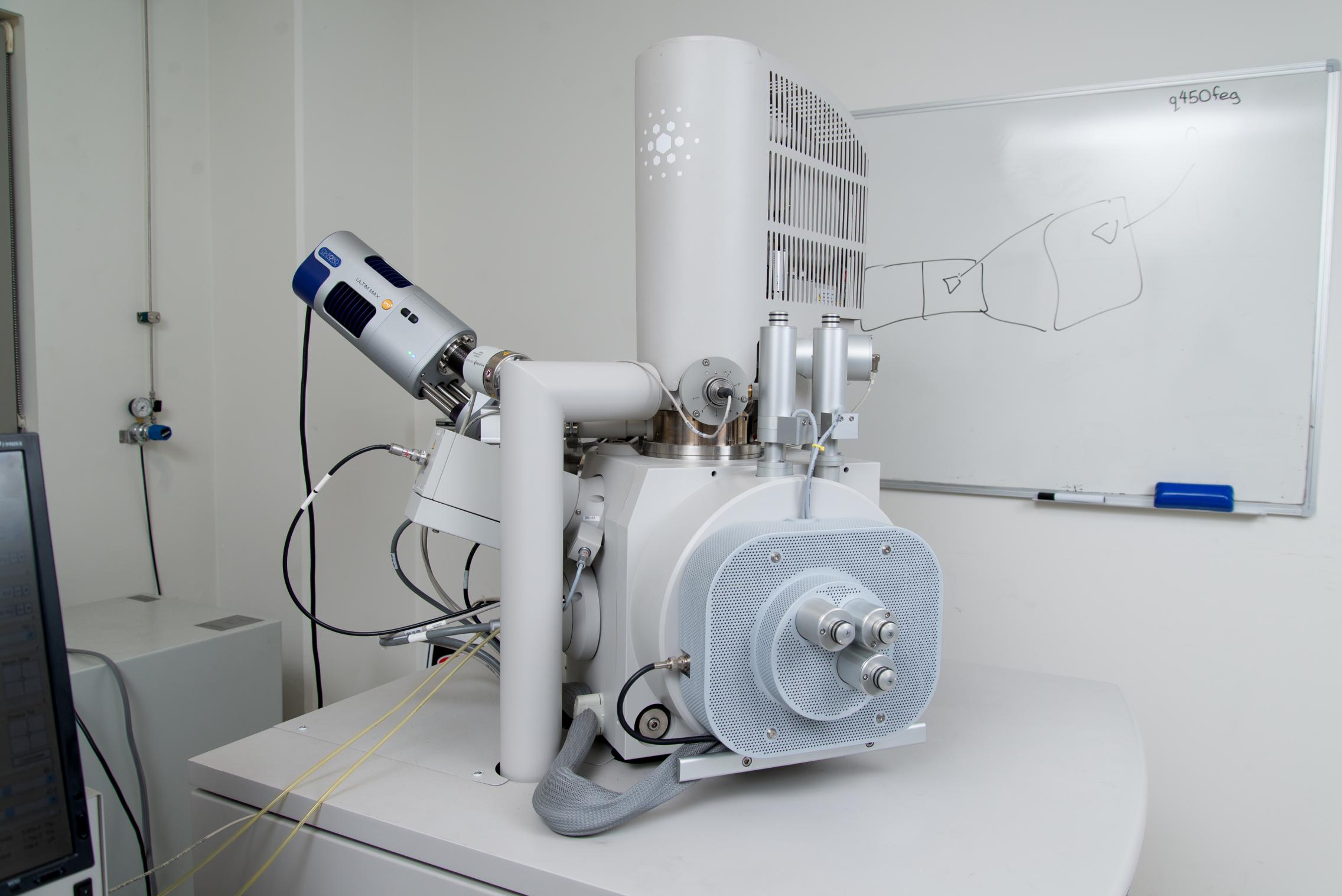
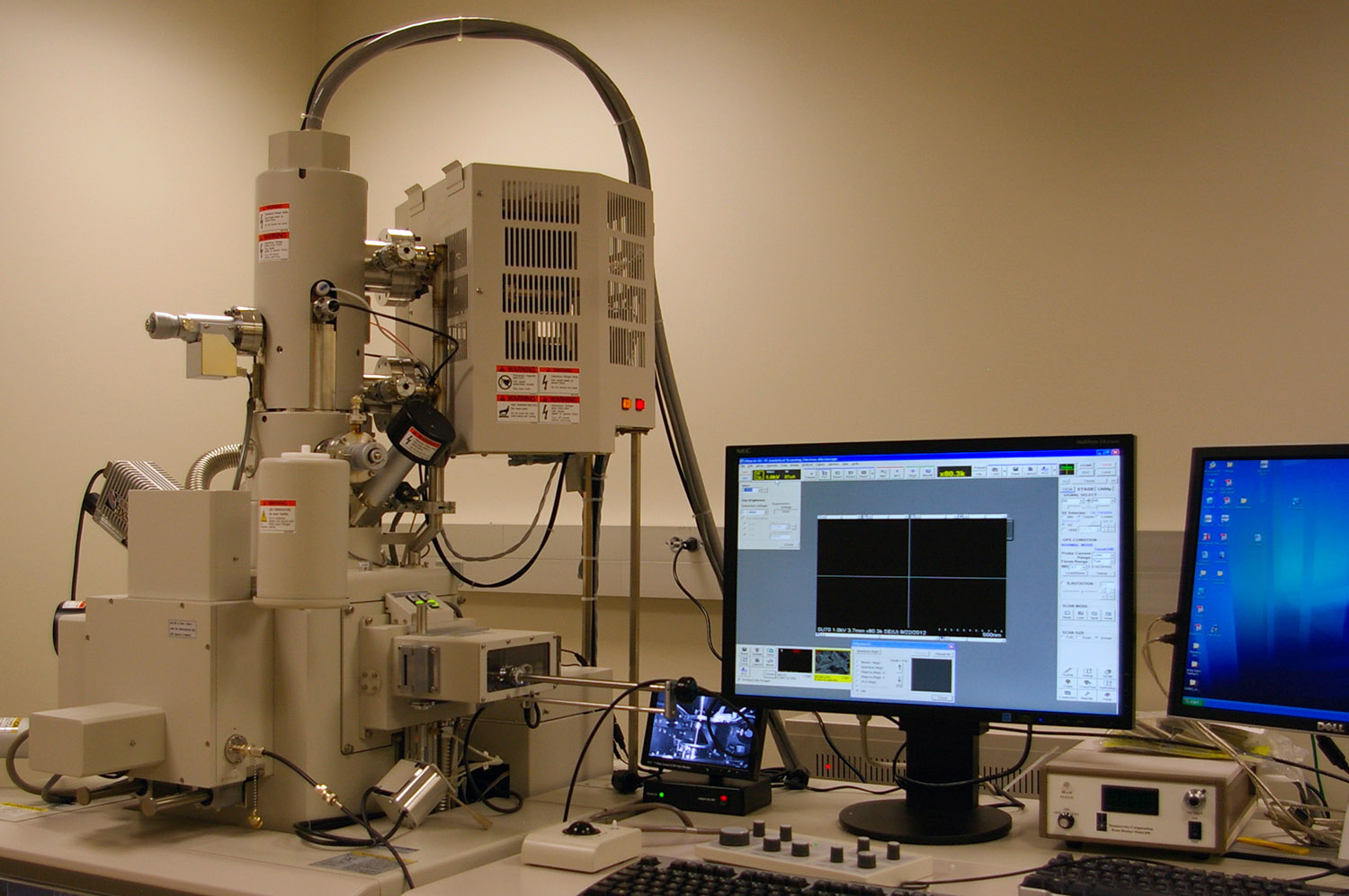
A typical SEM instrument, showing the electron column, sample chamber, EDS detector, electronics console, and visual display monitors. The scanning electron microscope (SEM) uses a focused beam of high-energy electrons to generate a variety of signals at the surface of solid specimens. The signals that derive from electron-sample interactions.
Field Emission Scanning Electron Microscope Dutco Tennant
Browse 1,200+ scanning electron microscope images stock photos and images available, or start a new search to explore more stock photos and images. Cancer cells vis - 3d rendered image, enhanced scanning electron micrograph (SEM) of cancer cell. Visual of overall shape of the cell's surface at a very high magnification.
Scanning Electron Microscope dpUNION
Scanning electron microscope is a classification of electron microscope that uses raster scanning to produce the images of a specimen by scanning using a.. botton: stomata on the leaf lower surface). Pictures reference - Wikipedia. Magnification Power of Scanning Electron Microscope. The magnification of an SEM ranges from 10X to 3,000,000X;
Scanning electron microscope introduced Scientist Live
Welcome to the exciting world of scanning electron microscope photography and video. Explore subjects from the ordinary to the extraordinary--from common objects and creatures to exotic animals and plants, microbiology, and high technology. This collection of photographic works spans 1974 to the present. View All Photo Categories »
Scanning Electron Microscope (SEM) Definition, Principle, Parts, Images Microbe Notes
Scanning electron microscope image shows SARS-CoV-2, 2019-nCoV, the deadly virus that causes COVID-19 emerging from the surface of cells cultured in the lab, 3d render. Using a scanning electron microscope and false colour techniques this image of a nettle leaf was established. The brown sections are the leaf veins, the green is the leaf cells.
FieldEmission Scanning Electron Microscope Nebraska Center for Biotechnology Nebraska
A scanning electron microscope (SEM) is a very high resolution microscope that allows one to see small things in very great detail. This is a quick overview on how to take pictures of a sample using one. Keep in mind that an SEM is a very delicate piece of equipment and should be used with great care.
JEOL 7600 Scanning Electron Microscope PNNL
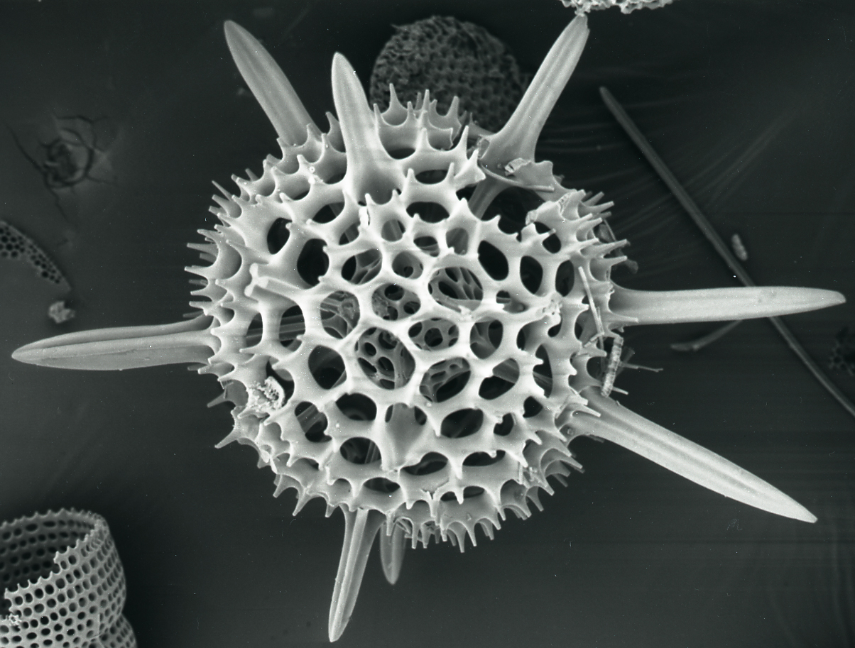
In scanning electron microscopy, a focused electron beam captures a high-resolution, grayscale image of a specimen by scanning its surface. Because the beam is sensitive to dust and water, this.

Field Emission Scanning Electron Microscope (FESEM) MERLIN iCRIM
Browse 2,068 authentic scanning electron microscope stock photos, high-res images, and pictures, or explore additional scanning electron micrograph or electron microscope micrographs stock images to find the right photo at the right size and resolution for your project.

Scanning Electron Microscope Tescan Vega 3 — Universidad de Monterrey
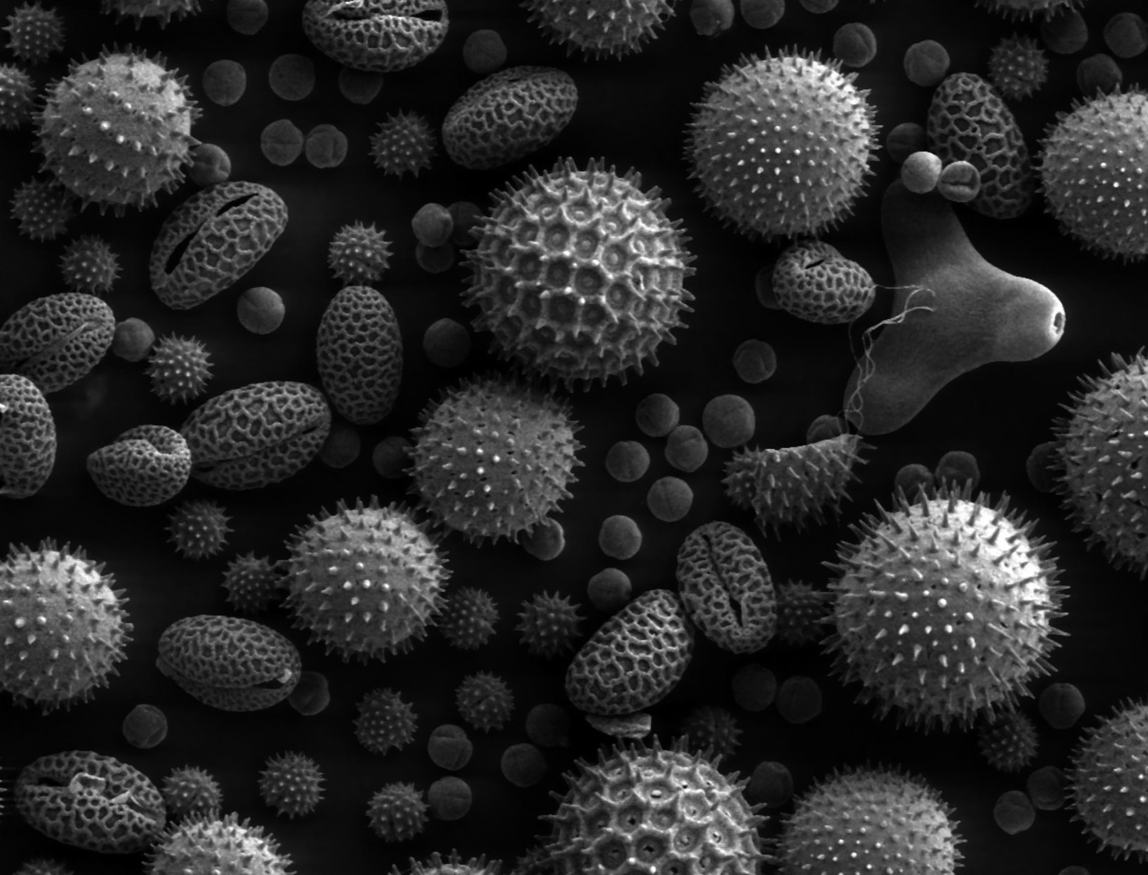
The microscopic world is an endlessly fascinating place, and thanks to technological advances over the last 90 years, we can now see things at incredibly high magnification through these electron microscope photos. Scanning electron microscopes (SEM) show us the invisible world of microorganisms by combining a variety of signals that are then.
Scanning Electron Microscopy Images Central Microscopy Research Facility
Coloured scanning electron micrograph (SEM), magnified x1500 when printed at 10cm wide. scanning electron microscope stock pictures, royalty-free photos & images. Fungus (Aspergillus niger), SEM. Aspergillus niger spores (reproductive cells). The fungus Aspergillus niger is a widely distributed saprophyte which grows on household dust, soil.